Melanoma skin cancer
Doctors sometimes use the term lesion to describe a finding on the skin. This means an area of skin that looks different from the surrounding area.
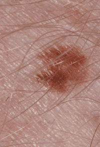
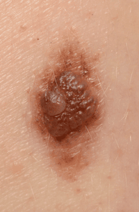
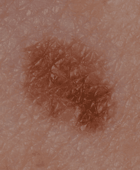
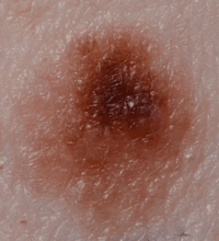
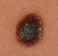
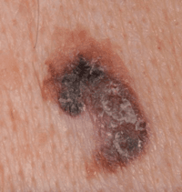
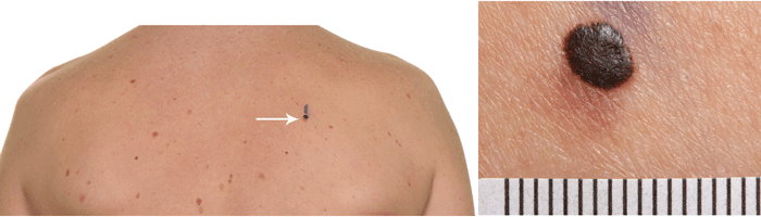
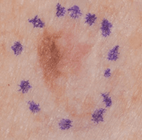
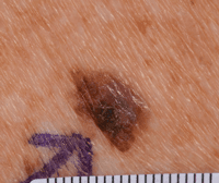
The following 2 examples contain 2 pictures. The first picture in each example is taken from a distance. The second picture from each example is a close up of the melanoma.
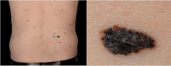
The blue markings in this picture below outline the area where the melanoma is. This is to show the surgeon the area they need to remove.
See your GP if you notice any changes that are not normal for you.
Find out more about seeing your GP if you are worried
The pictures above have been provided by the St John’s Institute of Dermatology at Guy’s and St Thomas’ Hospital. With special thanks to Dr Theodoulos Drousiotis for sourcing these photographs.
Last reviewed: 01 Apr 2025
Next review due: 01 Apr 2028
Symptoms include changes to a mole, freckle or normal patch of skin. Doctors use a checklist of signs to look out for. But it helps to know what your skin normally looks like.
You may be referred to a specialist if you have symptoms that could be due to melanoma skin cancer. This might be an urgent suspected cancer referral.
The risk of developing melanoma skin cancer depends on many things including how much ultraviolet radiation you get from the sun and your skin type.
Melanoma skin cancer starts in skin cells called melanocytes. You can get it anywhere on your skin including in a mole, on your palms, the soles of your feet and under your nails.
The stage of a melanoma skin cancer tells you how deeply it has grown down into the skin. It also tells you if it has spread elsewhere in your body and how far.
About Cancer generously supported by Dangoor Education since 2010. Learn more about Dangoor Education


Search our clinical trials database for all cancer trials and studies recruiting in the UK.
Connect with other people affected by cancer and share your experiences.
Questions about cancer? Call freephone 0808 800 40 40 from 9 to 5 - Monday to Friday. Alternatively, you can email us.